

Vision changes can range from a condition that needs monitoring to a retinal tear that requires urgent assessment. You will learn which symptoms need prompt attention, what to bring, how each examination and imaging test works, and how the findings shape treatment and follow-up.
Key takeaways
- Arrange urgent assessment for sudden flashes, floaters, or a curtain-like shadow.
- Bring your glasses, medicines, previous reports, and diabetes or blood-pressure records.
- Expect pupil dilation, vision checks, and tests such as OCT or retinal photography.
- Ask when treatment starts, what warning signs require urgent review, and when to return.
When Should You Arrange a Medical Retina Appointment?
New floaters, flashes, distorted straight lines, central blur, or gradual and unexplained loss of sight justify a medical retina appointment. Arrange routine assessment for persistent distortion, central blur, or vision changes that develop gradually.
| Symptom pattern | Appointment | Why |
|---|---|---|
| Sudden increase in floaters, flashes, curtain-like shadow, or abrupt vision loss | Urgent assessment, not a routine slot | These symptoms can indicate a retinal tear or detachment |
| Gradual central blur, distorted lines, or unexplained loss of sight | Routine medical retina appointment | The cause may involve the macula or retinal blood vessels |
At registration, staff record your name, contact details, referral information, and the affected eye. During history-taking, tell the clinician when symptoms began, whether they appeared suddenly or gradually, and whether they change when you read or look at a distance.
A medical retina specialist in Panchkula will ask about diabetes, blood-pressure control, pregnancy, allergies, blood thinners, previous eye injections, laser treatment, and other eye conditions.
The assessment may investigate diabetic retinopathy, diabetic macular edema, age-related macular degeneration, retinal vein occlusion, macular edema, or retinal inflammation. Take sudden symptoms seriously: waiting for a convenient routine appointment could delay treatment for a tear or detachment.
What Should You Bring and How Should You Prepare?
Bring your records and plan for temporary visual changes after dilation. Use this checklist before a retina examination in Panchkula:
1. Pack previous eye reports, OCT scans, fundus photographs, fluorescein angiography reports and any written diagnosis. Add diabetes records and blood-pressure readings if available.
2. Carry a current medication list with doses. Name every blood thinner, including aspirin, clopidogrel or warfarin, and write down the dates and names of previous eye injections or laser procedures.
3. Eat ordinary meals and take regular medicines unless the clinic gives different instructions. Confirm special preparation when fluorescein angiography, an injection or another treatment is planned.
4. Bring sunglasses. Dilating drops increase light sensitivity and blur near vision; arrange transport if you cannot drive safely afterward. Blurred vision can last several hours, and the duration varies between people.
5. Before fluorescein angiography, tell the team if you are pregnant, have allergies or kidney disease, or have reacted to injected contrast material before. This information helps them assess whether the test is safe and whether additional precautions are needed.
6. Keep your phone available for medication details and emergency contacts. If another person accompanies you, they can help with travel and instructions while your near vision remains blurred.
What Happens During the Eye Examination?
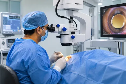
For a retina examination in Panchkula, the hands-on assessment usually moves from basic vision checks to a detailed view of the back of the eye. The order can change if early findings point to additional testing.
1. Visual-acuity testing measures each eye separately with an eye chart. You read it with your current glasses and without them, so the clinician can distinguish a focusing problem from reduced retinal vision.
2. Intraocular-pressure measurement checks the pressure inside each eye. The device used depends on the clinic and is usually quick.
3. A slit-lamp examination uses a focused beam and magnification to inspect the eyelids, cornea, lens and other structures at the front of the eye.
4. The clinician uses dilating drops when a view of the retina and vitreous is needed. Dilation is not optional for every retina assessment. The drops do not usually hurt, but they can sting briefly; near vision then becomes blurred and light sensitivity increases.
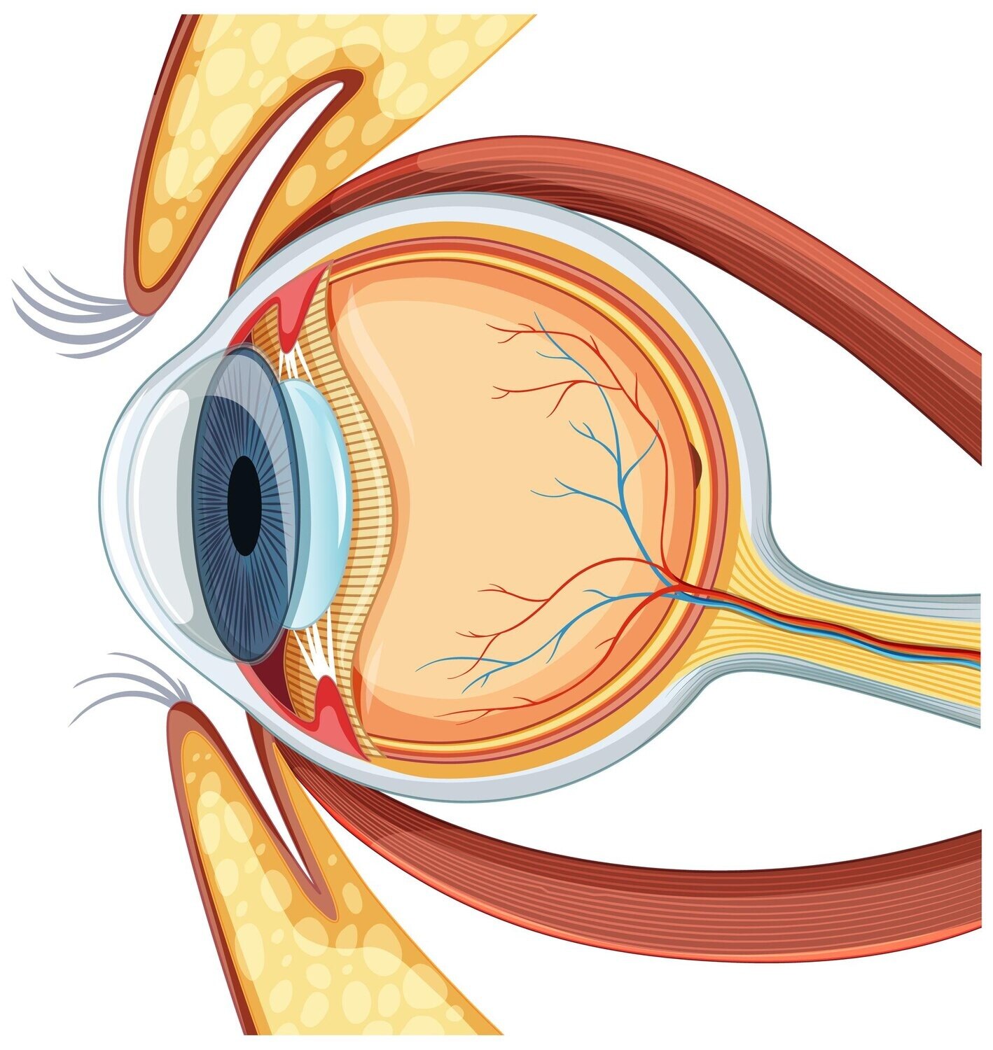
5. During the dilated fundus examination, the clinician examines the vitreous, optic nerve, macula and retinal blood vessels. This helps identify changes that a front-of-eye examination cannot show.
6. Drops need time to take effect, so the visit includes waiting before the retinal examination. OCT, photographs or other tests can extend the appointment; bring sunglasses and arrange transport if you cannot drive safely afterward.
Which Retina Tests Might Be Used and What Do They Show?
The suspected condition, clarity of the eye and findings from the first examination determine which retina tests you need. A retina examination in Panchkula does not make every investigation routine.
| Test | What it shows | What to expect and when it is used |
|---|---|---|
| Optical coherence tomography (OCT) | Cross-sectional images of the retina and macula, including fluid, swelling and structural changes | Quick, non-contact scanning with no injection; useful for detecting and monitoring macular disease |
| Fundus photography | The documented appearance of the optic nerve, macula and retinal blood vessels | A camera takes images, usually without discomfort, so the clinician can compare changes over time |
| Fluorescein angiography | Leakage and abnormal retinal blood vessels | Fluorescein dye enters a vein, followed by timed photographs; disclose pregnancy, allergies, kidney disease and previous contrast reactions before testing |
| B-scan ultrasonography | Information about the back of the eye when the retina cannot be seen clearly | Sound waves pass through the closed eyelid; painless for most patients and useful with cataract, vitreous haemorrhage or another opacity |
OCT is different from fluorescein angiography: it uses light, not injected dye, and does not show leakage in the same way. Fundus photography records appearance, while angiography traces dye movement through retinal vessels.
A medical retina service in Panchkula selects tests to answer a specific clinical question. An ultrasound scan can assess the hidden retina when an opaque lens or haemorrhage blocks the view, while OCT may be enough when the macula is visible.
How Are the Diagnosis, Treatment and Follow-Up Decided?
The clinician compares both eyes, then connects your symptoms and examination with OCT or angiography findings. This helps distinguish diabetic retinopathy, diabetic macular edema, age-related macular degeneration, retinal vein occlusion, inflammatory disease and other macular problems. Not every patient needs treatment at the first visit.
| Option | When it applies | Trade-off or follow-up |
|---|---|---|
| Observation with repeat OCT | Stable or uncertain findings | Delaying review can miss worsening fluid |
| Anti-VEGF injection | Macular edema, neovascular age-related macular degeneration or some proliferative diabetic retinopathy | Often requires a series of injections and monitoring |
| Laser | Selected leaking or ischaemic areas | Panretinal photocoagulation can reduce severe bleeding risk but affect peripheral or night vision |
| Steroid injection or implant | Inflammation or edema, especially when anti-VEGF is unsuitable or insufficient | Check eye pressure and cataract development |
For diabetes-related disease, glucose, blood-pressure and lipid control reduce progression risk; eye treatment cannot replace this medical management. A tear, detachment or vitreous problem needs a surgical retina referral rather than routine macular treatment.
After an injection, increasing pain, marked redness, worsening vision, or new floaters and flashes requires urgent contact. When comparing a medical retina service in Panchkula, ask whether it provides the imaging, specialist review, injection or laser planning, and follow-up schedule you need. Netra Eye Hospital is one local option for discussing that pathway.
A medical retina specialist in Panchkula can also explain how retina treatment in Panchkula fits your diagnosis and monitoring needs.
Related service
Clinical Services at Netra Eye Hospital provides comprehensive diagnosis, treatment, and long-term management of retinal diseases without surgery. View service → |
Frequently asked questions
When should you arrange a medical retina appointment?
Arrange an appointment for new floaters, flashes, distorted straight lines, central blur, or unexplained sight loss. Seek urgent assessment for sudden symptoms, a curtain-like shadow, or rapid vision loss.
What should you bring to a medical retina appointment?
Bring your glasses, current medicines, previous eye reports and scans, and records of diabetes or high blood pressure. Arrange someone to accompany you if dilating drops could affect your vision.
What happens during a retina examination in Panchkula?
The examination can include visual acuity testing, eye-pressure measurement, pupil dilation, and a detailed inspection of the retina. The specialist will ask about symptoms, timing, medical conditions and medicines.
Which retina tests might be used?
Optical coherence tomography shows cross-sectional retinal layers, while fundus photography records the retina’s appearance. Fluorescein angiography or other scans may be used when the specialist needs more information about blood flow or leakage.
How are retina treatment and follow-up decided?
The specialist matches treatment and review timing to the diagnosis, vision changes, scan findings and risk of progression. Options can include monitoring, injections, laser treatment, or referral for surgery when appropriate.